Simulation has a long history in medical education. As technology has evolved, simulation has evolved with it. If simulation is integrated into medical and graduate medical education curricula as the aviation industry has done, the licensing and board certification process as well as patient care and safety could be improved.
Introduction
Preventable medical errors are believed to be one of the largest causes of death in the United States (US), resulting in approximately 400,000 deaths per year.1 Likewise, iatrogenic injuries result in the disability of approximately 3.5 million patients per year in the US.1 These numbers are alarming. It is more shocking to consider that despite an abundance of new pedagogical methodologies and technologies, medical education has changed little in nearly 100 years and remains firmly rooted in apprenticeship.2-4 There is not only room for improvement, but also room to embrace tools that are currently available, namely simulation. Simulation as part of training and education has been successfully integrated into the curricula in other industries where errors have high consequences, notably aviation, to which medicine is often compared. Effective use of high-fidelity simulation has become a mainstay of commercial pilot education, so much so that when a commercial pilot flies an aircraft for the first time, it has a full load of passengers.⁴ Although simulation has been increasingly integrated into medical education, it has not occurred to the same extent as other industries such as aviation.
Origins of Simulation
With this in mind, it is surprising to learn that the application of simulation in medical education is not new. Ancient clay and stone models found across the globe were used to demonstrate clinical features of various diseases (figure 1).³ With the passage of time and advancement of technology, medical simulation has become more sophisticated. The first “modern” simulator, an interactive task trainer, was developed around 1700 by Gregoire and Gregoire, a father and son team in Paris, France, from a human pelvis and a dead baby.⁵ It was used primarily for teaching delivery methods to midwives and resulted in a demonstrable decrease in infant mortality.3,5 There is also ample documentation from the middle ages through modern times of the use of non-human animals in the development and teaching of surgical skills.⁶
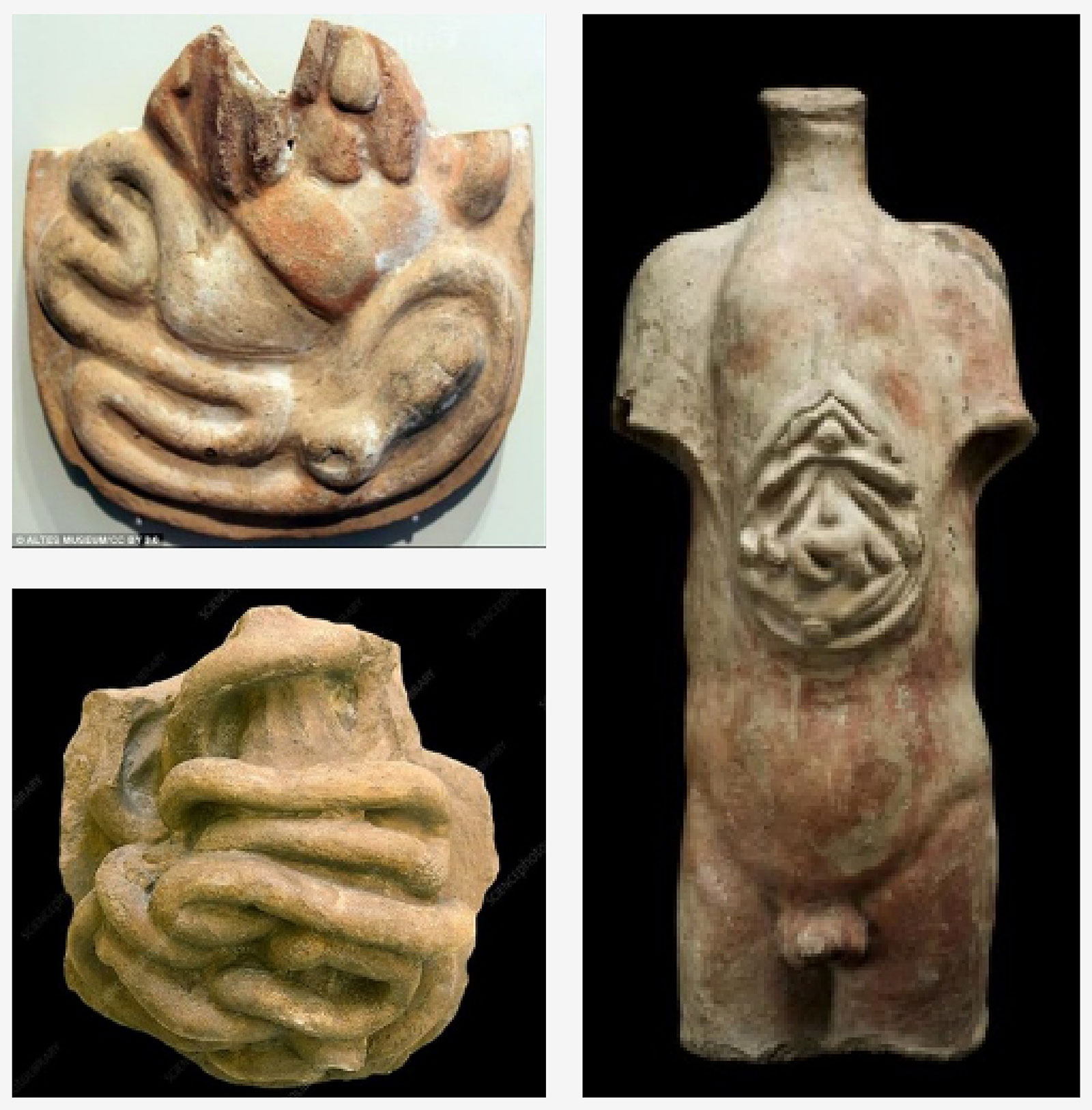
Figure 1: Ancient clay models of human anatomy recovered from sites throughout the Middle East, North Africa, and Central Asia. Left, anatomical votive offerings (Credit: Altes Museum, CC By 3.0. via Wikimedia Commons) Right, votive male torso, Roman, 200 BCE-200 CE. (Credit: Science Museum, London. Attribution 4.0 International (CC BY 4.0)
Development of Modern Simulation
The modern era of simulation in medical education began in the early 1960s after Dr. Peter Safar, working at Baltimore City Hospital, “rediscovered” and described “mouth-to-mouth” resuscitation.7,8 This work, and the prodding of a Norwegian anesthesiologist, Bjorn Lind, convinced Norwegian plastic doll and toy maker Asmund Laerdal to design and produce a realistic model of a human torso, allowing the application of Safar’s head tilt/chin lift to relieve airway obstruction and deliver mouth-to mouth rescue breaths.9 Later, at Safar’s urging, a spring mechanism was added inside the chest of Resusci-Anne® to allow for chest compressions.6,9,10 This was the origin of one of the most widely used CPR mannequins of the 20th century.⁵
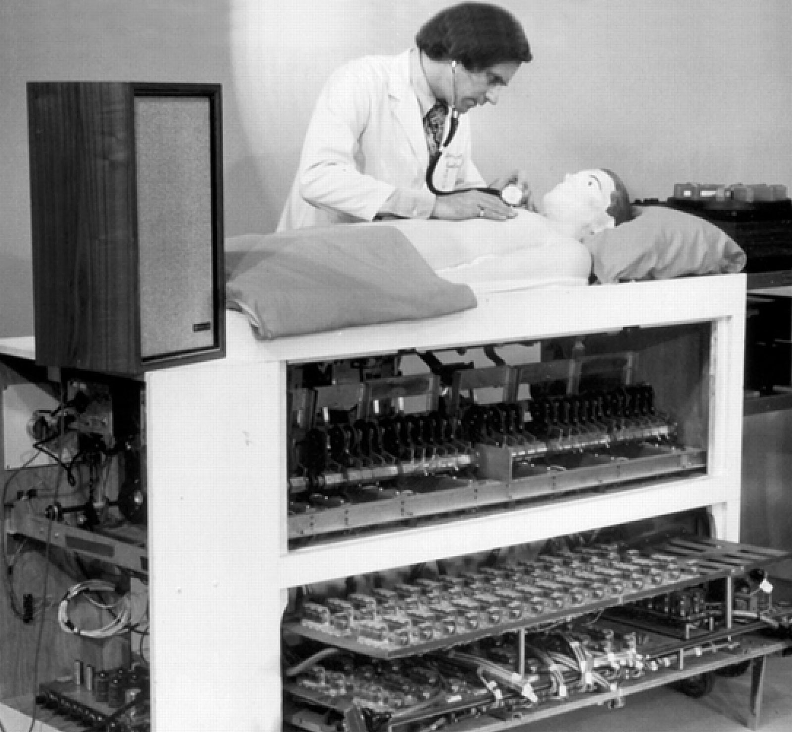
Figure 2: Michael Gordon demonstrating the original Harvey® cardiology simulator. Reused with permission from Cooper JB, Taqueti, VR. A brief history of the development of mannequin simulators for clinical education and training. Postgrad Med J. 2008; 84: 563–570.
Another major leap in simulation technology occurred in 1968 when Michael Gordon, MD, PhD, of the University of Miami presented Harvey®, the cardiology patient simulator (figure 2).⁶ Harvey® is capable of simulating almost any cardiac disease by presenting varying auscultatory findings, blood pressures, and pulse findings. It remains in service today at many medical schools, helping to teach physical diagnosis in cardiology.3,6,10
Resusci-Anne® and Harvey® are examples of the two major families of simulators in use today: task trainers, whose purpose is to teach a set of physical skills and diagnostic trainers, whose purpose is mainly to teach the interpretation of information. Task trainers have been developed for teaching everything from simple peripheral intravenous placement to laparoscopic surgical skills.5,6,10 Likewise, diagnostic trainers have been expanded to help medical trainees understand an array of patient information and presentations ranging from heart sounds to diagnostic imaging.5,6,10
Diagnostic trainers have further evolved to facilitate learning of patient interaction skills. In the early 1960s, Dr. Howard Barrows, a neurology resident at the New York Neurological Institute, made the astute observation that some patients, after repeated examinations by medical students and residents, would modify the neurologic findings on their examinations in response to repeated examinations by medical students and residents.3 When he graduated from residency and moved on to his own academic practice, Barrows began to train healthy actors to mimic various conditions; thus, in 1964, the standardized patient came into being.11,12
As computer hardware and software developed more rapidly throughout the 1980s and 1990s, the complexity and capabilities of simulators simultaneously evolved. The capability to simulate physiological states and responses to medications thereby providing real feedback to learners was developed. Anesthesiology simulation as a result began to take center stage. David Gaba and colleagues at Stanford University developed the Comprehensive Anesthesia Simulation Environment (CASE).® This tool advanced simulation beyond mere interaction with a mannequin to include a computerized waveform generator, which could produce all of the information typically found on patient monitors in the anesthesia environment.13,14 This development gave rise to the idea of simulators as environment trainers. Unlike task trainers or diagnostic trainers, environment trainers are not focused on learning skills or information but on the application of skills and information that the learner already possesses under a pre-established set of circumstances or conditions. This type of simulation immediately lent itself to applications such as anesthesia crisis resource management training.14,15
New Technologies in Simulation
As the capabilities of computers have continued to evolve, new technologies such as virtual reality, augmented reality, and mixed reality have been incorporated into simulation as well. Below, we provide definitions and examples of these terms.
Virtual reality is a fully immersive experience that tricks the user’s senses into thinking they are in a different environment separate from the real physical world. Using a head-mounted display or headset, the user can experience a computer-generated world of imagery and sounds in which digital objects can be manipulated using haptic controllers tethered to a console or PC. While in a virtual reality environment, interaction with the real world is limited. The most developed of these virtual reality simulators is the SimX® platform (San Francisco, CA),16 which allows multiple users to participate in the same simulation simultaneously. SimX® is one example of a platform that reacts to participants’ natural behavior and allows multiple users to be engaged in the same scenario (i.e., interacting with the same virtual patient and each other). As an example of the use of natural behavior when using this platform, if a participant were to pick up a virtual stethoscope in a virtual reality environment and apply it to the patient, the user can hear what they typically would through the stethoscope. Fundamental Surgery (FundamentalVR, London, UK),17 is a virtual reality platform designed for surgical training, also allows multiple users to interact with the same simulation and makes use of hand control devices, which mimic various surgical tools.
Augmented reality overlays digital information on real-world elements. Pokémon GO (Niantic, San Francisco, CA) is among the best-known examples. Augmented reality keeps the real world central in the simulation but enhances it with other digital details by the layering of new information that is not available without the computer additions thereby supplementing reality. Augmented reality allows for digital interaction with digital elements and physical interaction with real-world elements. One example is a platform made by GIGXR (Los Angeles, CA),18 which generates “holographic” patients in a real clinical environment. This system can be accessed using a head-mounted goggle system, which allows visualization of a virtual patient and displays his/her vital signs within the physical room where the user is located. The system can also be accessed using a smartphone or tablet, which uses the onboard camera to display the room and the virtual patient on the screen.
Mixed reality brings together real-world and digital elements. In mixed reality, the user interacts with and manipulates physical and virtual items and environments, using next-generation sensing and imaging technologies. Mixed reality allows the user to see and be immersed in the real world while physically interacting with both items in the real world as well as digital items. As a result, mixed reality breaks down barriers between real and imaginary. An example is the Heartworks® ultrasound simulation system19 by Intelligent Ultrasound (Cardiff, UK), which allows a user to place transthoracic and/or transesophageal ultrasound probes into a mannequin, manipulate the probe as would be performed at bedside, and explore how probe manipulation impacts the ultrasound image displayed on a computer monitor.
This system facilitates the acquisition of ultrasound probe manipulation skills and the examination of various pathologies, functioning as a combined diagnostic trainer and task trainer. Another example of a mixed-reality simulation system is the System of Modular Mixed and Augmented Reality Tracking Simulators (SMMARTS),20 which was developed at the University of Florida. SMMARTS is built around a core module that includes the tracking hardware and add-on modules that can be made to simulate essentially any desired anatomy.21 The physical module contains three dimensional-printed bony anatomy and a silicone gel or ballistics gel model of the surrounding soft tissue. The bony and soft tissue are modeled within the software environment. This allows the user to examine the tissue of interest and perform interventional procedures.21 Multiple modules have been developed for SMMARTS, including a spine for thoracic regional anesthetic procedures, a head for regional anesthetic procedures in the head and neck, a head for ventriculostomy procedures, a chest for internal jugular and subclavian central venous access, an arm for peripheral venous access, and a box for transrectal prostate examination and biopsy (figure 3).21-25
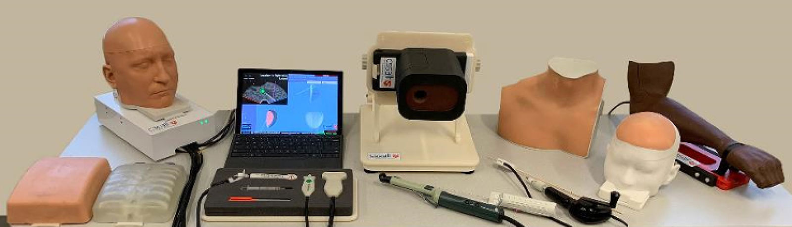
Figure 3: The current range of System of Modular Mixed and Augmented Reality Tracking Simulators (SMMARTS) simulator modules, including thoracic regional anesthesia, head and neck regional anesthesia, central venous access, peripheral venous access, ventroculostomy and prostate modules.
All of these technologies have been employed in various formats for medical education, primarily in the surgical and interventional care domains. They allow for ultrarealistic simulation of procedural skills without the need for a patient to be involved, for facilitation of anatomic diagnoses based on patient imaging data, or for complex surgical planning. Mixed-reality simulators offer multiple advantages because they are capable of simultaneously acting as diagnostic trainers, task trainers, and environment trainers.
One point of contention has been whether simulation practice can help improve patient safety. Although simulation training is gradually being adopted into medical curriculum, it still has not been widely practiced in many disciplines beyond advanced cardiovascular life support training or limited clinical crisis scenario exercises.26 Certain simple procedural tasks, such as central venous cannulation simulation, have indeed demonstrated a reduction of complications and improved patient outcomes.27 However, there remains a need for large prospective cohort studies to provide data that simulation training not only enhances medical procedure efficiency, but also improves patients safety.
Conclusions
As health care becomes more complex and clinical practice becomes more specialized, simulation is likely to continue to evolve to meet educational needs. We should expect virtual reality, augmented reality, and mixed-reality simulators to become increasingly more commonplace. Simulators are also likely to become more capable, integrating diagnostic, task, and environment trainers. Imagine a simulator mannequin that can generate waveforms and send them to anesthesia monitors, while simulating physical examination findings of a tension pneumothorax, allowing for bronchoscopic examination and endotracheal tube manipulation, central venous line placement, thoracocentesis, and chest tube placement, all using the same simulator tool. Not only would such tools be invaluable for medical education, but they would likely form the basis of a new paradigm for performance evaluation such as board certification, allowing for the examination of not only knowledge and judgement, but also physical skills. A broader adoption of simulation-based curricula into undergraduate and graduate medical education may have the potential to not only simplify evaluation, but also to improve the quality and safety of patient care.⁴
The authors would like to provide special acknowledgment to Leah Buletti for her editorial work with regards to this article.
Cameron R. Smith, MD, PhD, is assistant professor of Anesthesiology, Division of Acute and Perioperative Pain Medicine, Department of Anesthesiology, University of Florida College of Medicine, Gainesville, FL.
Yong G. Peng, MD, PhD, FASE, FASA, is professor of anesthesiology, associate professor of Surgery, and chief of the Cardiothoracic Anesthesia Division, Division of Cardiothoracic Anesthesia, University of Florida College of Medicine, Gainesville, FL.
Conflicts of interest: Cameron Smith, MD, PhD reports he is the inventor of the SMMARTS head and neck regional anesthesia simulator module but is not the patent-holder on the underlying technology. Yong G. Peng, MD, PhD, reports he has no conflicts of interest.
References
- James JT. A new, evidence-based estimate of patient harms associated with hospital care. J Patient Saf. 2013;9:122–128.
- Flexner A. Medical education in the United States and Canada. From the Carnegie Foundation for the Advancement of Teaching, Bulletin Number Four, 1910. Bull World Health Organ. 2002;80:594–602.
- Jones F, Passos-Neto C, Braghiroli OFM. Simulation in medical education: brief history and methodology. The Principles and Practice of Clinical Research. 2015;1:46–54.
- Dawson SL, Kaufman JA. The imperative for medical simulation. Proceedings IEEE. 1998;86:479–483.
- Buck GH. Development of simulators in medical education. Gesnerus. 1991;48 Pt 1:7–28.
- Cooper JB, Taqueti VR. A brief history of the development of mannequin simulators for clinical education and training. Postgrad Med J. 2008;84:563–570.
- Safar P. Ventilatory efficacy of mouth-to-mouth artificial respiration; airway obstruction during manual and mouth-to-mouth artificial respiration. J Am Med Assoc. 1958;167:335–341.
- Safar P, Escarraga LA, Elam JO. A comparison of the mouth-to-mouth and mouth-to-airway methods of artificial respiration with the chest-pressure arm-lift methods. N Engl J Med. 1958;258:671–677.
- Grenvik A, Schaefer J. From Resusci-Anne to Sim-Man: the evolution of simulators in medicine. Crit Care Med. 2004;32:S56–S57.
- Rosen KR. The history of medical simulation. J Crit Care. 2008;23:157–166.
- Barrows HS. An overview of the uses of standardized patients for teaching and evaluating clinical skills. AAMC. Acad Med. 1993;68:443–451; discussion 451–443.
- Barrows HS, Abrahamson S. The programmed patient: a technique for appraising student performance in clinical neurology. J Med Educ. 1964;39:802–805.
- Gaba DM, DeAnda A. A comprehensive anesthesia simulation environment: re-creating the operating room for research and training. Anesthesiology. 1988;69:387–394.
- Gaba DM, Lee T. Measuring the workload of the anesthesiologist. Anesth Analg. 1990;71:354–361.
- Gaba DM, DeAnda A. The response of anesthesia trainees to simulated critical incidents. Anesth Analg. 1989;68:444–451.
- SimX. Virtual Reality Health care Simulation. https://www.simxvr.com/. Accessed November 18, 2020.
- Fundamental Surgery. fundamentalsurgery.com. Accessed November 18, 2020.
- GIGXR. Available at: gigxr.com. Accessed November 18, 2020.
- Heartworks. Intelligent Ultrasound for smarter scanning. https://www.intelligentultrasound.com/heartworks/. Accessed November 18, 2020.
- The Center for Safety, Simulation and Advanced Learning Technologies. Augmented Reality & Mixed Simulation. https://simulation.health.ufl.edu/technology-development/augmented-reality-mixed-simulation/. Accessed November 18, 2020.
- Lampotang S, Bigos AK, Avari K, Johnson WT, Mei V, Lizdas DE. SMMARTS: An open architecture development platform for modular, mixed, and augmented reality procedural and interventional simulators. Simul Healthc. 2020 Sep 10. doi: 10.1097/SIH.0000000000000503. Online ahead of print.
- Bova FJ, Rajon DA, Friedman WA, et al. Mixed-reality simulation for neurosurgical procedures. Neurosurgery. 2013;73 Suppl 1:138–145.
- Hooten KG, Lister JR, Lombard G, et al. Mixed reality ventriculostomy simulation: experience in neurosurgical residency. Neurosurgery. 2014;10 Suppl 4:576–581; discussion 581.
- Robinson AR, 3rd, Gravenstein N, Cooper LA, Lizdas D, Luria I, Lampotang S. A mixed-reality part-task trainer for subclavian venous access. Simul Healthc. 2014;9:56–64.
- Sappenfield JW, Smith WB, Cooper LA, et al. Visualization improves supraclavicular access to the subclavian vein in a mixed reality simulator. Anesth Analg. 2018;127:83–89.
- Gaba DM. Simulation is a critical tool for advancing patient safety – avaliable to everyone regardless of location or resources. Online. APSF Newsletter. 2019;33:96–97.
- Barsuk JH, McGaghie WC, Cohen ER, O’Leardy KJ, Wayne DB. Simulation-based mastery learning reduces complications durig central venous catheter insertion in a medical intensive care unit. Crit Care Med. 2009;37:2697–2701.
