In patients who are anesthetized for surgery in the beach chair position, brain blood pressure can fall to levels below the brain’s ability to autoregulate. This can result in brain damage, but an understanding of cerebral autoregulation is essential to prevent this occurrence. Cerebral blood flow is at risk when cerebral blood pressure falls below approximately 70-80 mmHg. Prompt recognition and treatment of cerebral hypotension should eliminate the potential of brain damage in these patients.
See the original article online at: https://www.apsf.org/article/beach-chair-position-may-decrease-cerebral-perfusion/
 Many factors have decreased the incidence of anesthetic-related complications. Development of a cultural awareness and special emphasis on patient safety began with the acceptance of monitoring standards of care1 and has evolved since 1985 in many ways. The anesthesia-related mortality rate prior to 1985 was about 1:10,000 cases. Following publication of standards in JAMA, which we emphasized were applicable only to the Harvard Affiliated hospitals, public pressure led the American Society of Anesthesiologists (ASA) to adopt these standards verbatim within a few months.
The introduction of new technology, starting with pulse oximetry, followed by capnography, continued improvements in anesthesia machines and monitoring equipment, and safer drugs, etc., dramatically reduced anesthesia malpractice premiums and anesthetic-related mortality rates to one in several hundred thousand healthy ASA I and II patients.2-4
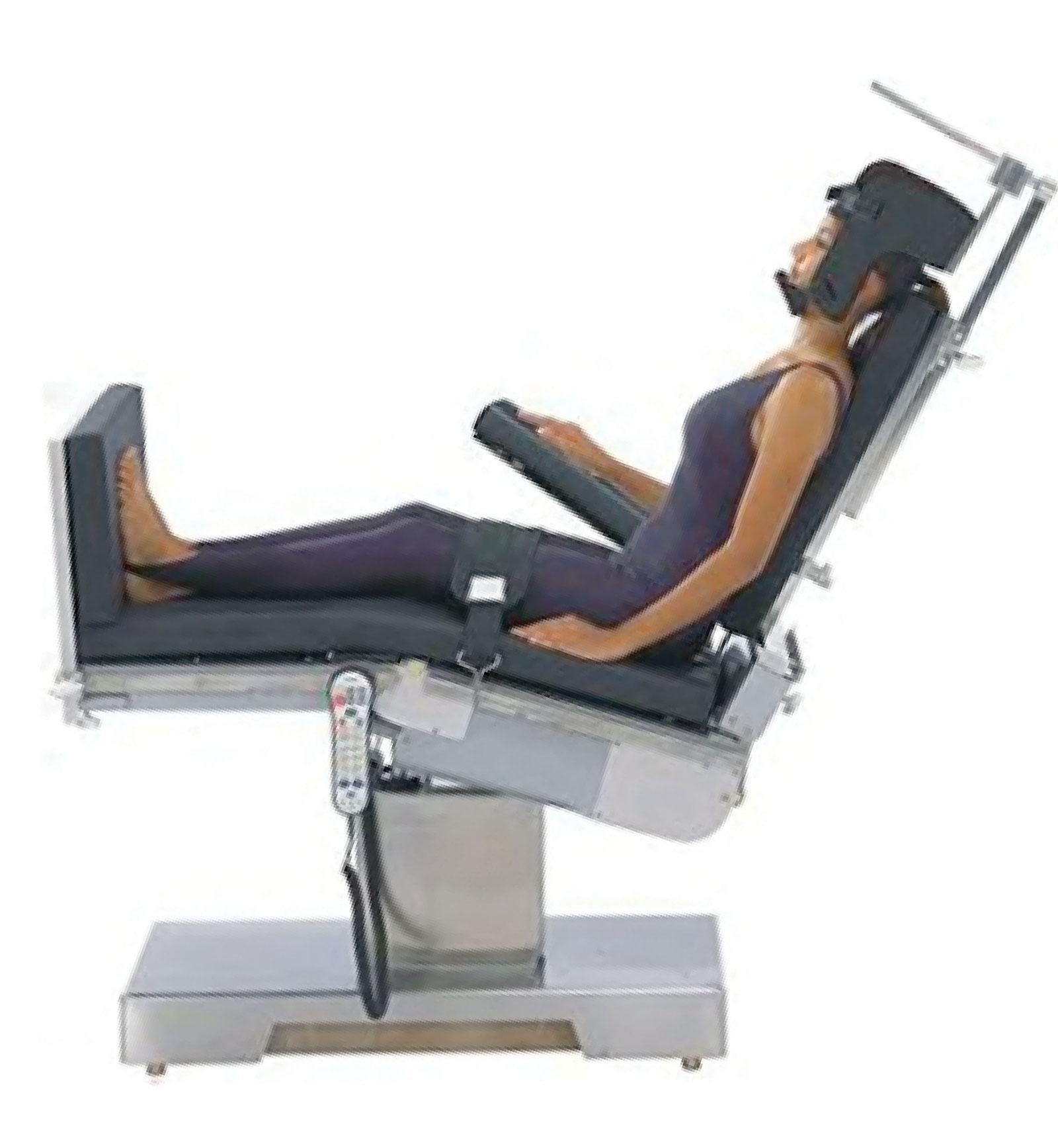
However, as exemplified by our original article,5 new problems will always arise, such as brain damage in healthy patients undergoing shoulder surgery in the beach chair position (BCP). Four cases came to my attention around 2000.5 Not one physician directly involved in the care of these four patients understood why the “stroke” occurred because, by their own admissions, the anesthesia professionals, surgeons, and subsequent neurology, cardiology and radiology consultants were unaware of the gravitational effects and hydraulic mechanism of the upright or BCP on cerebral perfusion. In short, cerebral perfusion pressure (CPP) fell to borderline or below threshold levels that ensured adequate cerebral perfusion.
For many well-known reasons, blood pressure (BP) almost always decreases during the early stages of general anesthesia in the BCP and is usually and safely reversed with fluid boluses and/or vasopressor infusion. When regional anesthesia to the shoulder is established prior to the operation, the surgical stimulus will be muted or absent, thus eliminating a potent way to counter the decreasing BP during surgery. Finally, many surgeons request/prefer that a mild degree of hypotension or even deliberate hypotension be maintained to reduce tissue swelling, limit bleeding and improve visualization in their surgical field.6,7 This leads to mean arterial pressures (MAP) that can be insufficient to sustain adequate cerebral blood flow (CBF) because, depending on the angle of the BCP and the height of the patient, the BP at the brainstem level will be approximately 20–40 mmHg lower than the BP as actually measured by cuff on the arm which is at the level of the heart.
As first described by Enderby et al. in 1954 regarding sitting craniotomies, for every inch in vertical height from the BP cuff placement on the arm to the brainstem, using the external auditory meatus (EAM), a surrogate for the brainstem level, one must subtract 2 mmHg of BP (or 1 mmHg per 1.25 cm.) to approximate the cerebral perfusion pressure (CPP).8 In 3 of the 4 cases I reported, and more I have reviewed, cuff systolic and diastolic BPs were usually in the 80s–90s/50s–60s measured at the arm/heart level and often lower. Therefore, MAPs at the brainstem would be about 20–40 mmHg lower, and at the level of the cerebral cortex, another 6–9 mmHg below that. Thus, MAPs in the brain would almost always be at or below the earlier established acceptable Lower Limit of Autoregulation (LLA), a MAP of about 50 mmHg.9
In the 1990s, studies by Drummond9 and others10 (Table 1) revised the LLA upward to account for the variable but incomplete vasculature in the Circle of Willis (found in 40–45% of cases), unpredictable collateral blood flow, and variations in the regional distribution of blood flow and cerebral oxygenation. Since the late 1990s, the range of the LLA has been revised upward and varies from 70–93 mmHg, with a mean value of 80 ± 8 mmHg.9,10 Recently, Brady et al. reported that the MAP with the most robust autoregulation during cardiopulmonary bypass in adults, obviously in the supine position, was 78 ± 11 mm Hg while the average LLA was 65 ± 12 mm Hg.11
Table 1: Lower limit of autoregulation in human studies10
| Investigators | LLA mean (mm Hg) |
| Strandgaard S. Br Med J. 1973;1:507–510. | 70 |
| Strandgaard S. Circulation. 1976;53:720–727. | 73 |
| Ohsumi H, et al. Resuscitation. 1985;13:41–45. | 81 |
| Waldemar G, et al. J Hypertens. 1989;7:229–235. | 93 |
| Schmidt JFG, et al. J Cardiovasc Pharmacol. 1990;15:983–988. | 85 |
| Larsen FS, et al. Stroke. 1994;25:1985–1988. | 79 |
| Olsen KS, et al. Br J Anaesth. 1995;75:51–54. | 88 |
| Olsen KS, et al. J Neurosurg Anesth. 1996;8:280–285. | 73 |
| Mean LLA for eight studies 1973 through 1996 | 80 ± 8 |
LLA = lower limit of autoregulation
The physical and hydraulic principles involved in the gravitational difference in MAP when using the upright or sitting position were well understood for decades. When sitting craniotomies were in vogue, it was standard practice, if monitoring intra-arterial BP, to zero the transducer at the height of the EAM. If only a BP cuff was used to monitor BP, correction for the vertical height from the cuff to the EAM was applied. When sitting craniotomies ceased being performed, this principle appears to have been forgotten or not taught to new anesthesia professionals.
In 2009, the APSF Symposium on Cerebral Perfusion in the Management of BCP Surgeries organized by Robert Stoelting, MD, resulted in most attendees agreeing that the mechanism of global ischemia (and I would add regional ischemia as well) had not been proven, but because the LLA has been revised upwards over the years, we should err on the side of caution when using deliberate hypotension or allowing patients to become hypotensive until we have better information.6 Obviously, this recommendation should be corrected to rule out the use of deliberate hypotension.
Anesthesia professionals cannot know the adequacy of circulation inside the brain because no routine clinical monitor exists which can monitor CBF, cerebral perfusion pressure (CPP) or cerebral tissue oxygenation during anesthesia in the BCP. By contrast for example, imagine an awake person sitting upright in a chair. For whatever reason, due to fear or fright, a sudden event, etc., their BP falls. Their first complaint would be lightheadedness, perhaps some nausea, or feeling faint. The first response would be to lay the person down, supine. This ensures, at least, that the CPP is the same as the BP at the heart and usually suffices to alleviate the distress. Unfortunately, the anesthetized patient cannot complain of these early symptoms as hypotension develops and affects the brain, so the anesthetic appears to proceed uneventfully. It is the responsibility of the anesthesia professional, to ensure as best they can, using indirect methods, that CPP and brain oxygenation are sufficient. During anesthesia, to ensure oxygenation, we first rely on knowing that the inspired oxygen concentration and oxygen supply from the anesthesia machine is sufficient. Next, to ensure the blood is well oxygenated, we rely on the pulse oximeter to monitor oxygen saturation and therefore know the blood going to the brain is well saturated. Then, by monitoring end-tidal CO2, we can maintain normal levels of CO2 to ensure that hypocarbia is not occurring, which would cause cerebral vasoconstriction. Finally, we use the BP measured at the arm to infer the CPP is high enough to move the well oxygenated blood through the brain. If the patient is supine, this assumption is reliable.
In the 2009 Symposium’s proceedings, published in the APSF Newsletter,6 current best practices recommendations for BP management in the BCP included the following: 1) Adjust BP in the BCP to account for the hydrostatic gradient; 2) Deliberate hypotension should be avoided in the BCP; 3) Maximum reduction from baseline BP should be no more than 30% with adjustment for any hydrostatic gradient in the BCP. In the opinion of myself and others, this recommendation should be changed. Instead, cuff BP should be maintained at or very near to the baseline awake BP when surgery is done in the BCP in order to protect the LLA.12,13 If necessary, BP should be restored to baseline by titrating fluids and vasopressors as needed.10
Scientific research was called for, and by now, many studies have been presented looking at ways to monitor cerebral oxygenation, regional cerebral blood flow, and jugular venous bulb oxygenation in relation to changes in BP. In a 2013 APSF article, Shear and Murphy reviewed the available studies on the impact of the BCP on cerebral perfusion.12 They wrote that until we know more about oxygenation and regional perfusion inside the brain, clinicians should remain aware of the danger of cerebral hypoperfusion in this patient population. In 2019, the same team extensively reviewed these and many newer studies.13 Twenty-two studies used various research tools to measure regional brain oxygen saturation, cerebral blood flow, and jugular venous oxygenation, and 68 studies looked at intraoperative management and outcomes. The authors summarized that there was often an imbalance of oxygen or CBF supply and demand during BCP surgery. However, an association between these variables of cerebral oxygenation and regional cerebral blood flow was not clearly shown. They concluded that in the absence of data generated within the patient’s brain, the safest approach toward perioperative BP management is to maintain MAPs close to baseline values throughout the procedure; wise advice. However, even if these studies of cerebral oxygenation and regional CBF had demonstrated a true cause and effect relationship between low BP and cerebral hypoperfusion or regional cerebral hypoxia, such research tools are not yet available for routine clinical monitoring. Perhaps in the future we’ll see the development of cost-effective, noninvasive monitors of CPP, CBF, and oxygenation using equipment derived from cerebral oximetry, near infrared spectroscopy, monitors of CBF, processed EEGs, or other new technologies. Until then, CPP must be maintained at extra safe levels, given what we currently know about the LLA versus how little we know about the adequacy of cerebral perfusion in each person’s brain during anesthesia.
Two very large studies of intra-operative hypotension (IOH) in patients undergoing a broad range of operations place the potential risk of the BCP to reduce cerebral perfusion into perspective. Monk et al. showed that about 5 minutes of BPs below the threshold limits for systolic BP of 70 mmHg, for MAP of 55 mmHg and for diastolic BP of 35 mmHg, with appropriate risk adjustment, was strongly associated with increased postoperative 30-day mortality from all causes.14 Similarly, Staplefeldt et al. extended this observation to find that when MAPs decreased progressively from 75 mmHg to 45 mmHg coupled with time of exposure to the IOH, the increase in all-cause 30-day postoperative mortality was also highly significant.15 A third study by Ahuja et al.16 examined myocardial and acute kidney injury in 23,140 patients undergoing noncardiac surgery, all of whom had intra-arterial BP measurements recorded at 1-minute intervals. When systolic BP fell below 90 mm Hg, and mean BP fell below 65 mm Hg, sustained for 5 minutes, significant and clinically meaningful associations were shown for myocardial and kidney injury. These three studies reinforce the concern that the risk of brain damage could also be increased when operating on patients in the BCP while not maintaining baseline BPs at the level of the brain. Why? Because the MAP at the brainstem (30–50 mmHg) and the cortex (20–40 mmHg) is lower, and the time of exposure to these very low BPs during surgery in the BCP is usually far longer than what was reported in these 3 studies.14-16 Thus, if a few minutes of decreasing MAPs towards 45 mmHg can increase postoperative 30-day mortality, and 5 minutes of decreasing MAPs below 65 mmHg can increase myocardial and kidney injury, it is reasonable to worry about the risk of brain damage when CPP decreases below 30–50 mmHg along with a one- or two-hour duration of cerebral hypotension as is common during shoulder surgery. Meanwhile, the anesthesia record will show a smooth and stable anesthetic since recorded cuff BPs at the arm/heart level seem relatively normal, having not been adjusted for the upright, BCP position.
Granted the outcome of brain damage is rare, as are many other catastrophic outcomes resulting from anesthetic complications. For example, malignant hyperthermia, or hypoxic encephalopathy, or death following failed intubation are rare outcomes, but enormous attention and resources justifiably have been and continue to be devoted to these topics and others. As Drummond et al. stated, “We cannot take assurance from the notion that at any given time “some” of the brain is not ischemic. It would be slim consolation to the devastated patients or their families to know that blood flow continued to some portions of the nervous system while disabling damage was evolving in others.”17
Knowing how autoregulation affects cerebral blood flow is critical in clinical practice because it leads us to gently react to mild decreases in BP in order to preserve cerebral perfusion. But, by knowing when the LLA (70–80 mmHg) is approached, thereby increasing the risk of cerebral ischemia as CBF falls in parallel with worsening hypotension, one must take into account the hydrostatic gradients and aggressively restore the patient’s BP to baseline at the arm/heart level. To paraphrase Lanier’s cautionary warning, this is consistent with our historic role as the vulnerable patient’s last homeostatic defense for avoiding brain damage during anesthesia and surgery.18
David Cullen MD, was formerly chairman in the Department of Anesthesia and Pain Medicine, St. Elizabeth’s Medical Center (Retired), professor of Anesthesiology Tufts University School of Medicine (Retired), formerly professor of Anesthesia and Critical Care Medicine, Harvard Medical School at Massachusetts General Hospital (Retired), Boston, MA
The author has no conflict of interest.
References
- Eichhorn JH, Cooper JB, Cullen DJ, et al. Standards for patient monitoring during anesthesia at Harvard Medical School. JAMA. 1986; 256:1017–1020.
- Eichhorn JH. Prevention of intraoperative anesthesia accidents and related severe injury through safety monitoring. Anesthesiology. 1989:70;572–577.
- Eichhorn JH, Hassan ZU. Anesthesia perioperative mortality and predictors of adverse outcomes. In: Lobato EB, Gravenstein N, and Kirby RR. (editors) Complications in Anesthesiology. 2007. 3rd ed. Philadelphia, Lippincott, Williams and Wilkins. pp 3–14.
- Eichhorn JH, Cooper JB, Cullen DJ et al. Anesthesia practice standards at Harvard: A review. J. Clin Anesth. 1988.1:55–65.
- Pohl A, Cullen DJ. Cerebral ischemia during shoulder surgery in the upright position: a case series. J Clin Anesth. 2005;17:463–469.
- Lee L, Caplan R. APSF Workshop: Cerebral perfusion experts share views on management of head up cases. APSF Newsletter. 2009;24:45–48. https://www.apsf.org/article/apsf-workshop-cerebral-perfusion-experts-share-views-on-management-of-head-up-cases/ Accessed August 12, 2020.
- Papadonikolakis A, Wiesler ER, Olympio MA, et al. Avoiding catastrophic complications of stroke and death related to shoulder surgery in the sitting position. J. Arthroscopic and Related Surgery. 2008;24:481–482.
- Enderby GEH. Postural ischaemia and blood pressure. Lancet. 1954;Jan 23: 185–187.
- Drummond JC. The lower limit of autoregulation: time to revise our thinking? Anesthesiology. 1997;86:1431–1433.
- Kirby RR, Cullen DJ. Complications of the beach chair position. In: Lobato EB, Gravenstein N, and Kirby RR. (editors) Complications in Anesthesiology. 2007 3rd ed. Philadelphia, Lippincott, Williams and Wilkins. 844–853.
- Brady KM, Hudson A, Hood R, et al. Personalizing the definition of hypotension to protect the brain. Anesthesiology. 2020;132:170–179.
- Shear T, Murphy G. Impact of the beach chair position on cerebral perfusion: what do we know so far? APSF Newsletter. 2013;28:18–20. https://www.apsf.org/article/impact-of-the-beach-chair-position-on-cerebral-perfusion-what-do-we-know-so-far/ Accessed August 12, 2020.
- Murphy G, Greenberg S, Szokol J. Safety of beach chair position shoulder surgery: a review of the current literature. Anesth Analg. 129:101–118.
- Monk TG, Bronsert MB, Henderson WG, et al. Association between intraoperative hypotension and 30 -day postoperative mortality in noncardiac surgery. Anesthesiology. 2015;123:307–319.
- Staplefeldt WH, Yuan H, Dryden DO, et al. The SLUS Score: A novel method for detecting hazardous hypotension in adult patients undergoing noncardiac surgical procedures. Anesth Analg. 2017;124:1135–1152.
- Ahuja S, Mascha EJ, Yang D. et al. Associations of intraoperative radial arterial systolic, diastolic, and pulse pressures with myocardial and acute kidney injury after noncardiac surgery. Anesthesiology. 2020;132:291–306.
- Drummond JC, Hargens AR, Patel TM. Hydrostatic gradient is important: Blood pressure should be corrected. APSF Newsletter. 2009;24:6. https://www.apsf.org/article/hydrostatic-gradient-is-important-blood-pressure-should-be-corrected/ Accessed August 25, 2020.
- Lanier W. Cerebral perfusion: err on the side of caution. APSF Newsletter. 2009 24:1–4. https://www.apsf.org/article/cerebral-perfusion-err-on-the-side-of-caution/ Accessed August 25, 2020.
