 Issue PDF
Our department recently received a warning from General Electric regarding the potential hazard of patients suffering burns during magnetic resonance imaging (MRI) with its equipment. I am not aware that any patient has actually been burned, even though the potential for burns exists. Why are we exposing patients to burns? Why does General Electric expect providers to assume some of the burden and liability of protecting patients from this potentially dangerous equipment-related issue? In the 1999 Institute of Medicine report,1 “To Err is Human,” the Institute of Medicine found that fixing “system problems” was better than depending on fallible providers to prevent injury: “Commonly, errors are caused by faulty systems, processes, and conditions that lead people to make mistakes or fail to prevent them.”1 General Electric’s warning is a “system problem” that should not be fixed by asking anesthesia professionals to insulate patients from the MRI bore during sedation or general anesthesia when patients are unable to respond to heat. Once the patient is in the MRI bore, it is difficult to visualize patient contact with the bore and prevent potential burns. An alternative solution and possibly a more appropriate one to this hazard is for General Electric to insulate the magnet bore so that burns are not possible.
Sincerely,
Donald H. Lambert, MD, PhD
Professor of Anesthesiology, Boston University School of Medicine and Anesthesiologist in the Department of Anesthesiology at Boston Medical Center, Boston, MA.
The author has no disclosure as it pertains to this article.
REFERENCE
- http://www.nationalacademies.org/hmd/~/media/Files/Report%20Files/1999/To-Err-is-Human/To%20Err%20is%20Human%201999%20%20report%20brief.pdf Accessed on June 1, 2018.
Reply:
As a medical imaging device manufacturer, we employ a “design for safety first approach” when developing magnetic resonance imaging (MRI) equipment. In some cases, however, MR imaging does require that MR operators follow well-established safety procedures to mitigate potential safety risks.
During an MRI exam, radiofrequency (RF) energy is used to excite protons within the body to create images from the anatomy of interest. The source of this energy is a whole-body RF transmit coil, which is a resonant electrical structure that uses time-varying current and voltage to generate the RF field. This device resides at the center of the MRI magnet and provides the mechanical structure for the patient bore.
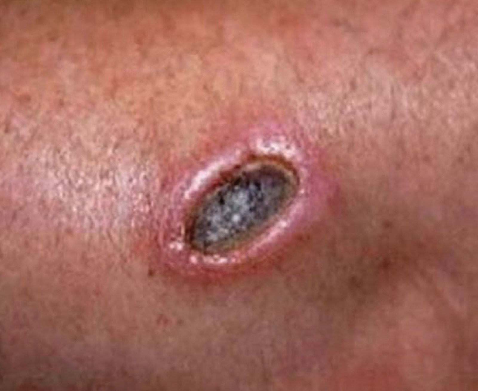
Figure 1. Depicts an example of an MRI radiofrequency-related burn.
Reproduced and modified with permission from GE Healthcare.
A consequence of the whole-body RF transmit coils’ operation is the induction of localized electric and magnetic fields within the patient’s body. As the anatomic structure of interest moves closer to the coil, the intensity of these fields can increase. Under certain conditions, the induced electric fields can result in warming of the tissue and in extreme cases, result in an RF burn (Figure 1). While uncommon, this represents a patient safety issue during MR imaging that must be taken into account during MR procedures by trained MR operators.
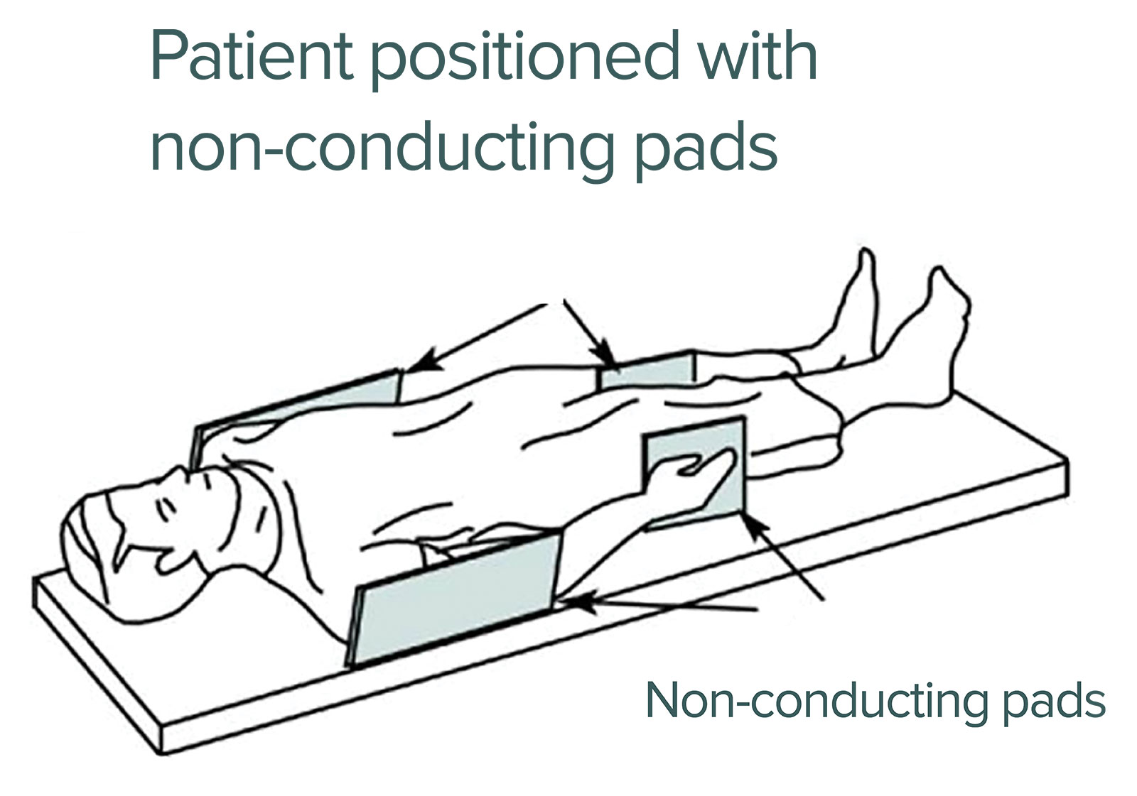
The MRI industry has developed safety procedures to address and mitigate the potential for RF burns during MR imaging. To prevent RF burns, every patient must be appropriately padded (Figure 2) using non electrically-conductive pads. These pads are placed between the patient’s skin and magnet bore. These pads should be a minimum of 0.25 inches thick and are furnished with every MR system (Figure 2).
|
For the issue described by Dr. Lambert, GE Healthcare has discovered a design issue with the RF body coil used in GE’s Discovery MR750w 3.0T scanner that may result in focal heating of the bore wall, above the 41 degree Celsius patient contact limit as defined by IEC60601-1 3.1, under certain conditions. The focal heating has been isolated to the circuit boards within the RF body coil assembly located at 0 (bottom), 90 (right), 180 (top), 270 (left) degrees on the table end of the patient bore (Figure 3).
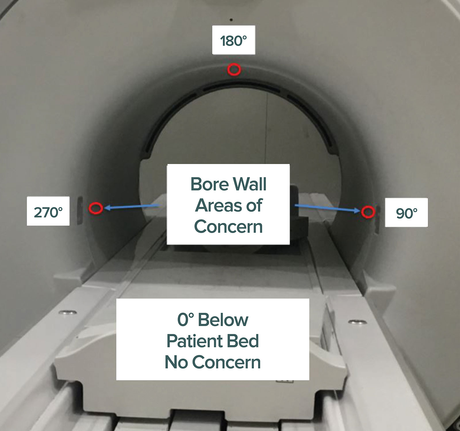
Figure 3. Depicts the particular areas where patients may be at increased risk for burn in MRI.
Reproduced and modified with permission from GE Healthcare.
In response, GE Healthcare has issued an URGENT MEDICAL DEVICE CORRECTION Ref# 60937 and will be correcting all affected Discovery MR 750W 3.0T products at no cost to the customer. While unrelated to RF burns, the safety instructions detailed in the letter are consistent with mandatory safety procedures described above and should be used as a mitigation for this focal heating issue. These instructions are designed to ensure customer awareness of the potential bore heating issue and remind MR operators of the required MR safety operating procedures.
Once again, thank you for providing GE Healthcare with the opportunity to reinforce the importance of following MR safety procedures.
Sincerely,
Bryan J. Mock, PhD
General Manager, Global 3.0T MR Segment
GE Healthcare – Imaging
The information provided is for safety-related educational purposes only, and does not constitute medical or legal advice. Individual or group responses are only commentary, provided for purposes of education or discussion, and are neither statements of advice nor the opinions of APSF. It is not the intention of APSF to provide specific medical or legal advice or to endorse any specific views or recommendations in response to the inquiries posted. In no event shall APSF be responsible or liable, directly or indirectly, for any damage or loss caused or alleged to be caused by or in connection with the reliance on any such information.