Introduction
 The act of positioning patients into a prone state is a routine event in operating rooms and procedural suites. Despite being common practice, physiologic changes and related injuries that accompany prone positioning are often overlooked by providers. This may be a result of lack of knowledge or a presumption of the routine. Regrettably, this approach may result in clinical decisions that result in an intraoperative crisis. This article aims to review potential injuries, highlight physiologic changes, and offer practical considerations when positioning and managing patients in the prone position.
Pressure Related Injuries
Injuries resulting from direct pressure often arise due to excessive strain on regions that are unaccustomed to sustained stress or load. Such pressure can either inflict harm directly or induce damage through diminished arterial inflow or obstructed venous outflow resulting, respectively, in ischemia or edema. Friction from slight movements of the body can affect the head and face or extremities and cause skin damage, including blistering. It is critical to meticulously safeguard vulnerable areas such as the eyes, nose, cheekbones and cheeks, forehead, chest, arms, breasts, genitalia, pelvis (including superior iliac spines), knees, and feet.1 Shoulders, being susceptible to dislocation during positional shifts, also require careful attention.1 Ensuring thorough protection can significantly reduce the risk of developing pressure sores and nerve injuries during extended medical procedures.2,3
Nerve ischemia, which can result from excessive stretching or direct pressure leading to microvascular compression, is another potential risk. Vulnerable nerve regions include the superior orbital nerves, lingual and buccal nerves (often strained due to unintentional jaw retraction caused by tension between masseter muscles), the phrenic nerve and recurrent laryngeal nerve (potentially impacted by over extension or rotation of the neck), the brachial plexus, the ulnar nerve, and the lateral cutaneous nerve of the thigh.1,2 Risk factors may include improper positioning of the limbs, long cases, inadequate padding, anatomic variations, hypotension, diabetes, hypothermia, and malnutrition.2
Physiologic Changes
Ocular
Ocular injury can occur due to either direct or indirect pressure. Improper patient positioning may inadvertently place weight and pressure on the eye, leading to ischemia.2 Also, the prone position can reduce intraocular perfusion by reducing venous outflow and increasing intraocular pressure, effectively lowering intraocular perfusion. Furthermore, the prone position can also elevate intra-abdominal pressure, reduce preload and mean arterial pressure (MAP), and further reduce intraocular perfusion. These physiologic changes may increase the likelihood of optic nerve ischemia, ischemic optic neuropathy, and central retinal artery occlusion.1,3 Visual loss following anesthesia and surgery in the prone position may be associated with increased surgical duration, large blood loss, and administration of large volumes of crystalloid IV fluid.4,5
The prone position is a risk factor for potential blindness in patients with closed angle glaucoma due to increased IOP, reduced blood flow, and eye compression.2 Due to the potential for a reduction of aqueous humor outflow in the prone position, intraocular pressure may increase, resulting in optic nerve injury. In addition, general anesthesia can decrease tear production. Coupled with incomplete eye closure, patients can be at increased risk for corneal injury and irritation.2
Head and Neck
In the prone position, there is a risk of increased intracranial pressure and reduced cerebral blood flow; this may lead to intracranial vessel distension. These changes could be especially concerning for patients with known or unknown intracranial space-occupying lesions.
Patients positioned prone are also susceptible to vascular injury, potentially affecting the head, neck and chest. Over-rotation of the neck may significantly restrict carotid or vertebral arterial blood flow and impede venous drainage. The prone position also increases hydrostatic pressure and can cause dependent edema. These alterations could result in complications such as stroke, tongue swelling, tracheal compression, or oropharyngeal and glottic edema.1,3,7 Dependent edema may also manifest as significant facial and scleral edema in addition to lingual edema.6,7 Such occurrences may present as unexpected airway difficulties or require delayed extubation. Reintubation may become prohibitively challenging with what was once a normal airway now presenting as a can’t intubate, can’t ventilate, scenario.
Pulmonary
Pulmonary function may improve in the prone position by improvements in functional residual capacity, improved ventilation to perfusion (V/Q) matching, and by raising arterial oxygen tension. While chest wall and lung compliance remain unchanged, it is important to note that intrathoracic pressure and peak airway pressure can be elevated in the prone position.1,2
Obesity and sleep-related breathing disorders can considerably increase the perioperative risk of cardiac complications.5 Given the heightened risk of increased pulmonary vascular resistance linked with the prone position, echocardiography to identify both the existence and severity of systolic and diastolic dysfunction may be considered in high-risk patients.6
Cardiovascular
Cardiovascular alterations related to disturbances in preload, afterload, and contractility are among the key factors to consider when positioning a patient in a prone position. Research has shown that the prone position can lead to an average drop of 24% in cardiac index, largely due to a decrease in stroke volume.1 Patients in this position may also experience tachycardia and an increase in peripheral vascular resistance. The occurrence of increased pulmonary vascular resistance and right ventricular strain due to acidosis can create hemodynamic instability in patients with a history of heart failure, pulmonary hypertension, or restrictive/obstructive lung disease.6
The prone position also increases the likelihood of mediastinal compression, as the right ventricle can be compressed against the sternum.1 The prone position can also increase intrathoracic pressure, reduce inferior vena cava filling, reduce atrial filling, and reduce left ventricular compliance with subsequent decrease in cardiac output. The use of bolsters (specially designed support devices or padding used to maintain a patient’s position during surgery) in the setting of chest and abdominal positioning is crucial. Local compression of anterior chest wall or abdomen may catastrophically reduce right ventricular function or inferior vena cava preload respectively. Patients with scoliosis, pectus excavatum, or recent cardiothoracic surgery may be at heightened risk.1 In addition, anesthetic agents can have a profound effect on cardiopulmonary physiology. Volatile anesthetics and propofol are known to decrease systemic vascular resistance, alter heart rate, and decrease cardiac contractility (due to direct myocardial suppression).
Risk factors for cardiovascular collapse in the prone position include massive blood loss, hypothermia, fluid shifts, cardiac comorbidities, venous air embolism, and anatomic deformities (e.g. thoracic lordosis or pectus excavatum).1,3 Extreme care should be taken with patients with right ventricular dysfunction, pulmonary hypertension, or in patients who are preload-dependent or highly sensitive to elevations in pulmonary vascular resistance.6 These patients may not tolerate the right ventricular strain associated with prolonged prone positioning.5 A thorough discussion should be undertaken to determine the safety of the prone position. This patient subset may be at a heightened risk of myocardial infarction and cardiac arrest when transitioned to the prone position.
Intra-abdominal
The prone position may potentially trigger abdominal compression, particularly in patients with obesity.2 This can result in decreased arterial inflow and venous outflow to the visceral organs, which has led to reports of pancreatitis and hepatic ischemia.1,2 It is of paramount importance to be careful in minimizing direct pressure on the abdomen. The risk of venous bleeding, due to venous engorgement as well as postoperative thrombotic complications, can be heightened by intra-abdominal venous compression.
Extremities
The likelihood of harm can vary based on a particular position and duration of the procedure. Some positions, such as bending of the hips and knees, can decrease arterial blood circulation, thereby increasing the chances of conditions like limb compartment syndrome, rhabdomyolysis, and subsequent renal failure.1
Clinical Recommendations
Preoperative
A thorough and focused preoperative exam is warranted for all patients scheduled in the prone position. In addition to patient history, airway examination, and preexisting neurologic deficits, the preoperative discussion should also include the projected duration of the procedure and proposed patient positioning. The patient’s capacity to handle the prone position should be carefully evaluated.
It should be noted that patients with anatomical deformities that may make positioning challenging or hazardous may also have syndromes that predispose them to requiring procedures in the prone position, such as spine surgery or percutaneous nephrolithotripsy. In addition to thorough discussion, it may be appropriate to attempt the desired position out of the operating room in advance of the day of surgery, with the patient in street clothes, attempting to position the patient in the desired position. Also, once it has been demonstrated that the position is possible for a given patient, consider photographing details of the position for upcoming operations. In our facility, positioning details for particularly high-risk cases are documented in the preoperative record, and images of the positioning specifics are uploaded to the electronic medical record.
If cardiac testing is warranted, guidelines for perioperative assessment are provided through the American College of Cardiology (Washington, D.C.) and the American Heart Association (Dallas, Texas). A cardiac work up not only provides risk assessment for ischemic heart disease, but also provides an assessment of right ventricular function, pulmonary hypertension, and valvular disease.6
If prone positioning is not suitable, consider whether the lateral or supine position would be more appropriate. If the prone position is inevitable, particularly in high-risk patients, advanced hemodynamic monitoring should be considered, including but not limited to central venous catheter, arterial catheter and even echocardiography along with the potential use of inotropes and vasopressors.
Intraoperative
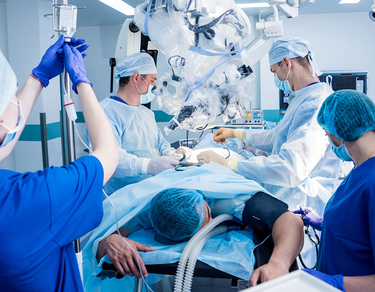
When positioning the patient, care should be taken to ensure proper padding. The eyes, mouth, neck, and all pressure points should be checked periodically to avoid any pressure-related injuries. The team responsible for positioning the patient should ideally consist of 5-6 members, including the surgeon or the proceduralist. If necessary for anesthetic planning, arterial and venous access should be placed prior to changing position if the arms are to be tucked.
Cervical inline stabilization should be maintained during position change, and the patient’s head and neck should be placed in a neutral position. Care should be paid to securing the endotracheal tube to prevent dislodgement during position changes or during the operation, especially if the head is in pins or a halo.1 Tape may not be ideal for securing the endotracheal tube during a long prone case, especially if the patient has facial hair. Unless the planned operation is on the head or neck, flat tracheostomy ties can be used to secure the endotracheal tube. If the operation involves head or neck, it may be necessary to suture the endotracheal tube to a tooth, the jaw, or place the endotracheal tube nasally and secure to membranous nasal septum. Gravity should not be allowed to act on the endotracheal tube or circuit. Providers should also be aware that neck flexion may result in the endotracheal tube becoming main-stemmed into the right or left bronchus.
The positioning of the arms depends on the type of surgery. Arms should be moved independently of each other to prevent shoulder joint injury upon initial positioning and case completion; extra padding should be placed to minimize the risk of ulnar nerve injury and to ensure the axilla is free from tension to reduce the risk of brachial plexus injury. Also, consider placing the arms slightly anterior to the shoulders in the coronal plane, with the arms less than the individual patient’s full extension at the elbow joint to protect the brachial plexus as well as to avoid biceps tendon pain.8
Following immediate placement of the patient into the prone position, the vital signs should be monitored. The bed/gurney should remain available in the room until the patient is noted to tolerate prone positional hemodynamically. In case the patient does not tolerate the change of position, they can be turned supine and resuscitated quickly. Once hemodynamic stability is noted following change of position, the bed/gurney can be removed.
At the end of the case, an evaluation for facial, lingual, and glottic edema should be made. Keeping the head of the bed slightly elevated during the case may minimize edema.1 This final evaluation may help guide disposition planning and ultimately guide the decision for extubation or the need for a higher level of care or remaining intubated until edema involving critical airway anatomy has resolved.
Conclusion
Complications arising from the prone position are recognized, but frequently underestimated. It is important for anesthesia professionals to understand the typical pressure-related injuries and physiological alterations that can occur in standard cases where the prone position is utilized. It is also crucial for providers to conduct a thorough preoperative history, assess the patient’s capacity to tolerate the prone position, and consider alternative positions for high-risk individuals. When the prone position is unavoidable, a meticulously planned strategy should be implemented in partnership with the surgeon or proceduralist.
Taizoon Q. Dhoon is an associate professor of Anesthesiology at the University of California, Irvine, California, USA
Shermeen Vakharia is a professor of Anesthesiology at the University of California, Irvine, California, USA
Evan Villaluz is an assistant professor of Anesthesiology at the University of California, Irvine, California, USA
Debra E. Morrison is a professor of Anesthesiology at the University of California, Irvine, California, USA
The authors have no conflicts of interest
References
- Edgcombe H, Carter K, Yarrow S. Anaesthesia in the prone position. BJA: British Journal of Anaesthesia. 2008;100(2):165–183. doi:10.1093/bja/aem380
- Kwee MM, Ho YH, Rozen WM. The prone position during surgery and its complications: a systematic review and evidence-based guidelines. Int Surg. 2015;100(2):292-303. doi:10.9738/INTSURG-D-13-00256.1
- Feix B, Sturgess J. Anaesthesia in the prone position. Continuing Education in Anaesthesia Critical Care & Pain. 2014;14(6):291-7.
- Lee LA, Roth S, Posner KL, Cheney FW, Caplan RA, Newman NJ, Domino KB. The American Society of Anesthesiologists Postoperative Visual Loss Registry: analysis of 93 spine surgery cases with postoperative visual loss. Anesthesiology. 2006 Oct;105(4):652-9; quiz 867-8. doi: 10.1097/00000542-200610000-00007. PMID: 17006060.
- Anesthesia Patient Safety Foundation. APSF Newsletter. 2020;35:85.
- Carabini LM, Koski TR, Bebawy JF. Perioperative Management for Complex Spine Fusion Surgery. Anesthesiology. Published online December 4, 2023. doi:10.1097/ALN.0000000000004744.
- Walsh A, et al. Association of Severe Tongue Edema with Prone Positioning in Patients Intubated for COVID-19. Laryngoscope. 2022;132(2):287-289.
- Graverson, JA, D Morrison, J Cho, A Kaplan, C Abdelshehid, A Lusch, M Liss & J Landman. Ureteroscopy: patient positioning and room setup. Cur Clin Urol 2013.
